
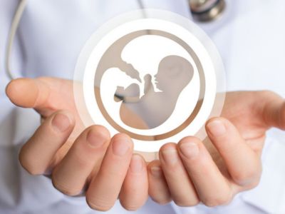
We, Delhi IVF & Fertility Research Centre, are delivering our admirable services over the globe, being situated in the heart of the capital of India-New Delhi. We are dedicated to offering the highest success in IVF treatments at a cheap cost for IVF in Delhi, India. We came into presence in 1993 and centered on giving good, passionate, moral and most advanced & specialized treatment to couples suffering from and finding answers as well to the problem of infertility..


Challenges are just opportunities in disguise. View all country!
20,000+
IVF Babies

50-81%
Success Rate

65+
Countries

12,000+
Successful Fertility Surgeries


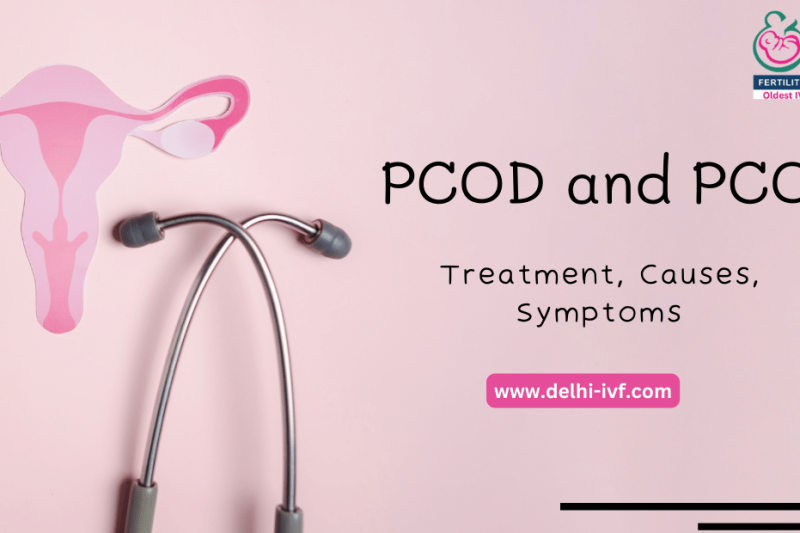
Female Infertility
Delhi-IVF is an ideal female infertility treatment clinic in Delhi, India which may bring the joy in the lives of infertile patients

Male Infertility
In humans, 40-50% of infertility is due to some problem with the male. It affects about 7% of all men and it is due some deficiency

IVF-ICSI
ICSI/IVF is advancement over IVF to improve the chances of having a pregnancy by making sure the egg is fertilized by

MESA/TESA
Some of the causes of male infertility are non availability of normal viable sperms in the ejaculate for IVF treatment.

Multiple Failed IVF
Repeated IVF failure/Recurrent Implantation Failure refers to the failure of clinical pregnancy. Here is the Treatment of IVF failure.

Unexplained Infertility
Unexplained Infertility refers to a condition where the reason of infertility in the male/female or both partners is not known.

Cryopreservation
Cryopreservation is the advanced process of storing tissues, cells or organs at very minimum and low temperature to preserve

Donor
Some women can’t use their own eggs because of non-availability of good eggs or low ovarian reserve, or premature ovarian failure

Endoscopy
An endoscopy is referred to a nonsurgical procedure of looking inside the body. An endoscope is an instrument containing a flexible tube with lenses

Altruistic Surrogacy
Surrogacy in India is a procedure in which a woman carries the baby of another couple/single parent up to the full term. It is done with the legal agreement between the two parties.
IVF with Donor Services
IVF Centers in India – IVF treatment or In Vitro Fertilization is fertilization in a “test tube” and the process is carried outside the women’s body in

PRP / Ovarian Rejuvenation
Platelet-rich plasma is a concentrate of platelet-rich plasma protein acquired from whole blood. It is prepared by a method
Delhi IVF Frequently Asked Questions
IVF FAQ
As such, IVF treatment is considered to be the best treatment for dealing with infertility. When we talk about good IVF treatment then yes India has extremely reputed and experienced IVF centres and skilled specialists, making it a good option for IVF treatment, which make India one of the Highest IVF success rate producers, with a lower IVF cost than any other country.
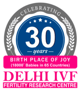
Delhi IVF Fertility & Research Centre is the pioneer of modern Fertility treatments since 1993 having served more than 25000 international patients from 65 countries over the last 3 decades. With latest technology and recent advancements, the IVF experts at the center ensure the people seeking healthcare are provided with the best.
International FAQ
Success rates vary globally, but countries like Denmark and Sweden India have high success rates. But yes, European countries are expensive. However , India is also emerging to provide the best & successful IVF treatment with the highest IVF success rates and the most affordable price.
In India, many clinics documenting success rates exceeding 45-80% and Delhi IVF Fertility & Research Center is one of the oldest IVF clinic for Assisted Reproduction, offers world-class IVF treatment in India following all the advanced procedures, techniques, and equipment, which have been making a significant mark to its high success rate.
In India IVF started in the year 1993 and currently India has emerged as a best country to get IVF treatment done. With the best & well experienced IVF specialists, India provides very advanced IVF treatments not only for the people in India but also for international patients. There are many clinics who offer a 45-80% IVF success rate which is much higher than other countries.
Delhi IVF & Fertility Research Center is one of the oldest clinics which had its first IVF clinic in 1993 and was established in the same year by Dr. Anoop Gupta. Dr. Anoop Gupta, an IVF expert for reproductive medicine & technology. DIFC has its head office in New Delhi and is a reputed IVF clinic for its IVF success rate, Personal Care, IVF Counselling, Quality Control. DIFC has helped more than 25000+ couples from the across the world 65 countries in the last 3 decades. It’s been one of the most trusted IVF centres for so many years and enjoys a great reputation in the general public in India and overseas.
The time it takes to conceive through IVF varies; it may take several cycles before achieving a successful pregnancy. IVF isn’t a singular treatment but a series of procedures. On average, an IVF cycle spans approximately 2.5-4 weeks from the initial consultation to the transfer stage. However, it’s important to note that while the general path remains consistent for all patients, what differs is how your body responds at each phase.

Delhi IVF & Fertility Research Centre, for Assisted Reproduction, offers world-class IVF Treatment in India following all the advanced procedures, techniques, and equipment, which have been making a significant mark to its high success rate.
We have been recognized & appreciated by many global reproductive societies like in the USA, Europe, South East Asia, and many others. Our systems and facilities are at par with the latest IVF Delhi techniques & technologies worldwide.
Reasons to choose Delhi IVF & Fertility Center


Our primary goal has been to provide Fertility information to all cities and countries. We ensure to keep sharing all valuable IVF in India news.


Delhi IVF's Media Coverage


 Your Journey to Parenthood Begins with us!